
Previous Topic
Chemistry
Biology

Home

Math
Physics

Next Topic
Previous Topic
Chemistry
Biology
Home
Math
Physics
Next Topic
The human body is a biological machine composed of systems; these are groups of organs that work together to produce and maintain vital functions.
In total, there are 11 systems in the human body.
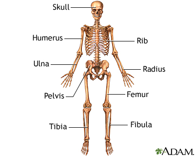
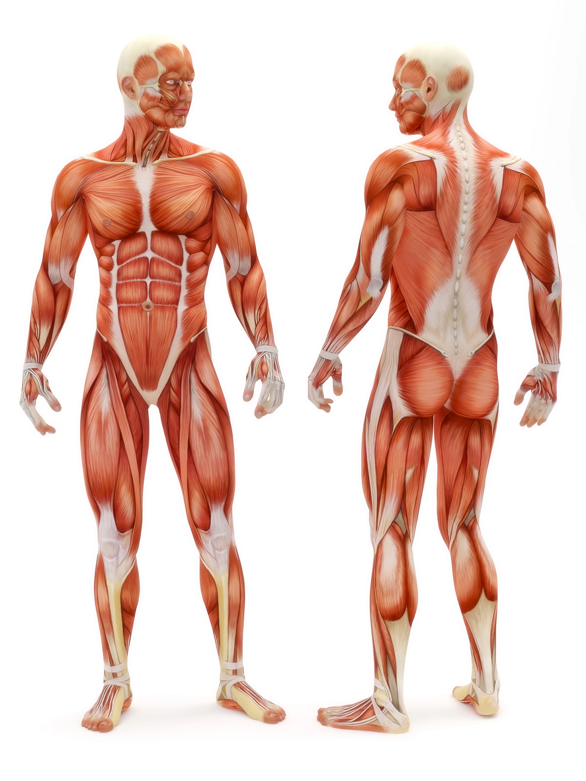
The skeletal system is composed of bones and cartilage. There are two parts of the skeleton: the axial and the appendicular. The axial skeleton consists of the bones of the head and trunk. The appendicular skeleton consists of the bones of the limbs, as well as the shoulder girdle and pelvic girdle.
There are a total of 206 bones in the adult human body. The place where two bones fit together is called a joint. Joints are supported by cartilage and reinforced by ligaments. Some functions of the skeletal system are mechanical support, movement, protection, blood cell production, calcium storage, and endocrine regulation.
The components of the skeletal system are tailored to the functions of the body parts they support. Thus, the anatomy of bones, joints, and ligaments is studied topographically, such as the bones of the head, neck, thorax, abdomen, and upper and lower extremities.
The muscular system consists of all the muscles in the body. There are three types of muscle: smooth muscle, cardiac muscle, and skeletal muscle.
Smooth muscle is found within the walls of blood vessels and hollow organs like the stomach or intestines.
Cardiac muscle cells form the heart muscle, also called the myocardium.
Skeletal muscles, on the other hand, attach to the bones.
Of the three types of muscles, only skeletal muscles can be consciously controlled and allow us to move our bodies. The other two are regulated by the autonomic nervous system, which is completely unconscious.
Viewed through a microscope, skeletal and cardiac muscle fibers are arranged in a repeating pattern, giving them a striped appearance, which is why they are called striated muscles. In contrast, smooth muscle does not contain repeating sarcomeres, so it is not striated muscle.
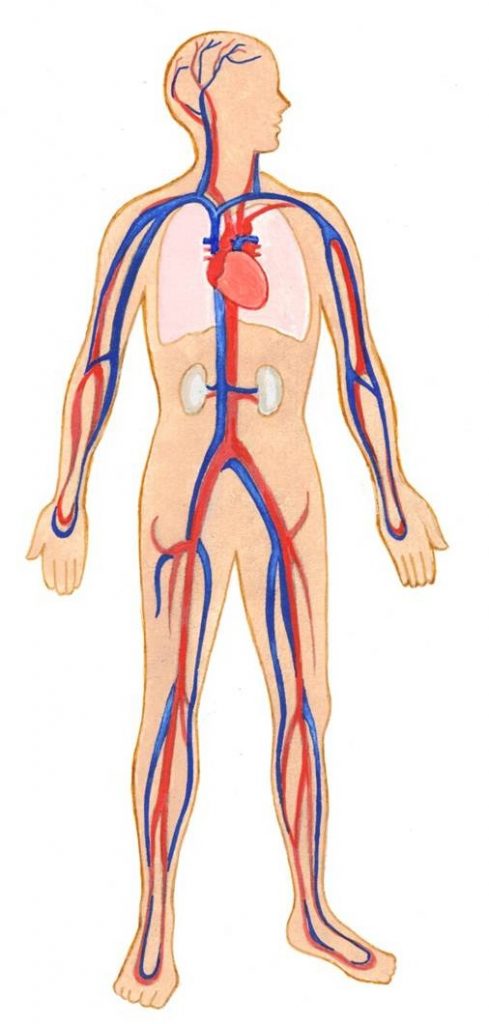
The cardiovascular system consists of the heart and the circulatory system of blood vessels. The heart is composed of four chambers: two atria and two ventricles. Blood enters the heart through the upper chambers, the left and right atria, and exits through the left and right ventricles. Heart valves prevent backflow of blood.
The heart acts as a two-way pump. The right side of the heart pumps deoxygenated blood into the pulmonary circulation, where the blood is reoxygenated. Meanwhile, the left side of the heart simultaneously pumps oxygenated blood into the systemic circulation, distributing it to peripheral tissues. The heartbeat is controlled by the cardiac conduction system.
The circulatory system, also called the vascular system, consists of arteries, veins, and capillaries. Together they make up the network of blood vessels that act as conduits to transport blood throughout the body. Blood leaves the heart through arteries, which progressively narrow to become smaller arterial vessels called arterioles. The arterioles terminate in a network of even smaller vessels called capillaries. Gas and nutrient exchange occurs across the walls of the capillaries.
Small veins, called venules, arise from the capillaries and gradually increase in size as they approach the heart until they become veins. There is some histological difference between arteries and veins, but their main functional difference is reflected in the direction in which they carry blood: arteries carry blood from the heart to the periphery, while veins carry blood from the periphery to the heart.
There are three separate circuits in the circulatory system.
1. The pulmonary circulation that carries blood between the heart and lungs.
2. The coronary circulation that supplies the heart muscles.
3. The systemic circulation that carries blood to the rest of the body.
The main arteries of the systemic circulatory system are the aorta and its branches; While the main representatives of the veins are the superior vena cava and the inferior vena cava.
The main functions of the cardiovascular system include the transport of oxygen, nutrients, and hormones throughout the body through the blood, as well as the removal of carbon dioxide and other metabolic waste products.
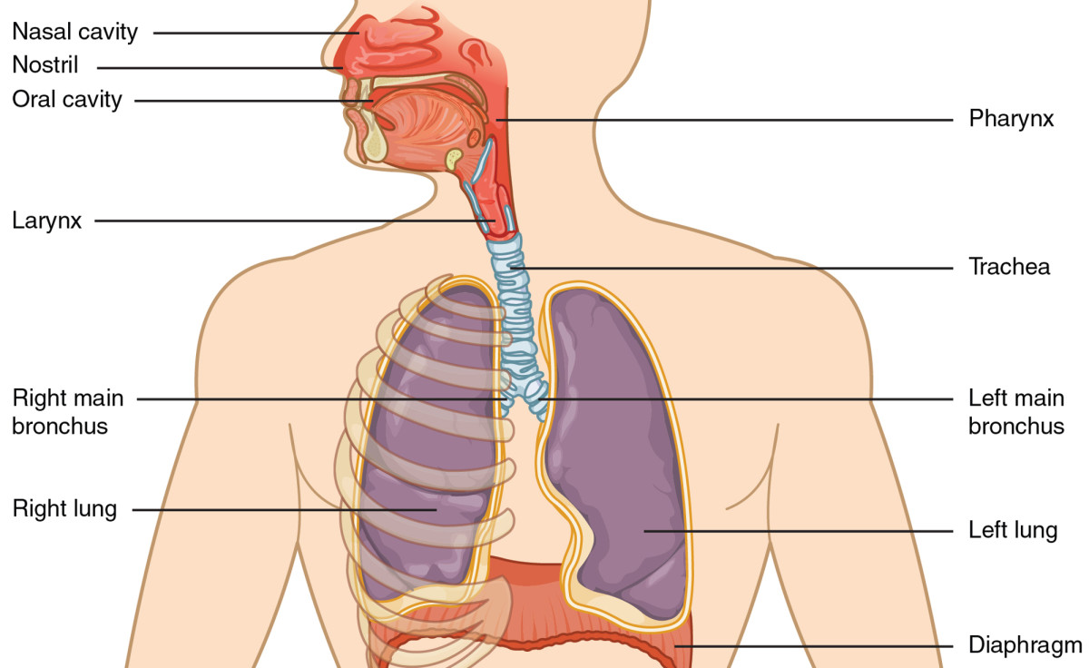
The respiratory system consists of several organs: the nasal cavity, pharynx, larynx, trachea, bronchi, bronchioles, and lungs (alveoli).
Together, the nasal cavity and pharynx are called the upper respiratory system, while the remaining named organs comprise the lower respiratory system.
The organs of the respiratory system, with the exception of the alveoli, function to convey air to the lungs with the help of the respiratory muscles (primarily the diaphragm and intercostal muscles).
Once air enters the lungs, it continues to the alveoli (the site of gas exchange) and interacts with blood transported by the pulmonary circulation. This is where carbon dioxide is removed and oxygen enters the blood. So the main function of the respiratory system is to bring oxygen into our body and remove carbon dioxide.
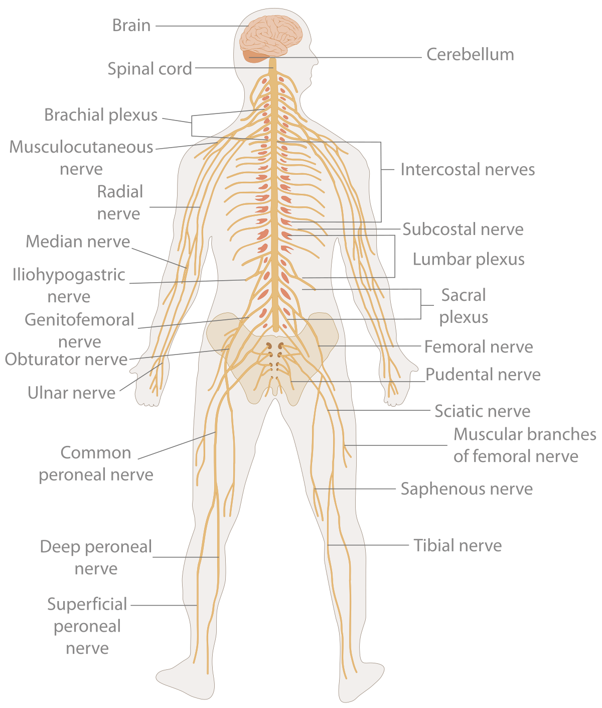
The nervous system is responsible for how we interact and respond to our environment by controlling the functions of our organs in the different systems.
The organs of the nervous system are the brain, spinal cord, and sensory organs. These are interconnected by neurons, which transmit nerve signals throughout the body.
Morphologically and topographically, the nervous system is divided into the central nervous system (CNS) and the peripheral nervous system (PNS). Functionally, the nervous system is considered to be two parts: the somatic, or voluntary, nervous system and the autonomic, or involuntary, nervous system.
The central nervous system is defined by the fact that it receives information from the environment and generates instructions accordingly, thereby controlling all activities of the human body. This information is two-way, meaning it enters and leaves the CNS, and its transport is through the peripheral nervous system.
The CNS consists of the brain and spinal cord. The brain is located within the neurocranium and is composed of the cerebrum, cerebellum, and brainstem (pons of the brain and medulla oblongata). The central portions of the CNS are filled with spaces called ventricles, filled with cerebrospinal fluid (CSF). The spinal cord is located within the vertebral column. The vertebral canal extends through the central portion of the spinal cord. This canal is also filled with CSF and communicates with the ventricles of the brain.
The CNS is composed of neurons and their processes (axons). Thus, it forms two parts: The gray matter, which is made up of neuronal cell bodies, is found in the cerebral cortex and the central portion of the spinal cord. The white matter, which is made up of axons, which combine to create neuronal pathways. The gray matter is where instructions are generated, while the white matter is the medium through which these instructions are transported to reach the organs.
The peripheral nervous system is defined by its conduct of information from the CNS to target organs, and from these organs to the CNS. It consists of nerves and their ganglia. The nerves that carry information from peripheral sensory organs (e.g., the eye, tongue, nasal mucosa, ear, and skin) to the CNS are called sensory, or afferent, ascending nerve fibers. The fibers that carry information from the CNS to the periphery (muscles and glands) are secretory, or motor, efferent nerve fibers.
A ganglion is a collection of neural tissue outside the CNS, composed of neuronal cell bodies. Ganglia can be either sensory or autonomic. Sensory ganglia are associated with spinal nerves and some cranial nerves (V, VII, IX, X).
Peripheral nerves emerge from the CNS. There are 12 cranial nerves that arise from the brain, and 31 pairs of spinal nerves that emerge from the spinal cord. Cranial nerves are numbered from I to XII, according to their exit from the skull (from anterior to posterior). The spinal nerves are divided into 8 cervical, 12 thoracic, 5 lumbar, 5 sacral, and 1 coccygeal nerve, depending on the vertebral level from which they arise. In certain regions of the body, peripheral nerves interconnect, creating neural networks called plexuses.
Some important plexuses are:
Cervical plexus (C1-C4) - innervates the back of the head, some cervical muscles, the pericardium, and the diaphragm via the greater auricular, transverse cervical, lesser occipital, supraclavicular, and phrenic nerves.
Brachial plexus (C5-T1) - innervates the upper extremities with nerves such as the median, ulnar (ulnar), radial, musculocutaneous, and axillary nerves.
Lumbar plexus (L1-L4) - innervates the muscles and skin of the abdomen and pelvis, as well as the thigh muscles via the iliohypogastric, ilioinguinal, genitofemoral, lateral cutaneous, obturator, and femoral nerves.
Sacral plexus (S1-S4, with some branches from L4, L5) - innervates the muscles and skin of some regions of the pelvis, posterior thigh, lower leg, and foot via the following nerves: gluteal, sciatic, posterior cutaneous of the thigh, pudendal, piriformis nerve, obturator internus nerve, and quadratus femoris nerve.
The somatic nervous system (SNS) and the autonomic nervous system (ANS) are divisions of the peripheral nervous system, with information carried through the cranial nerves and spinal nerves.
The somatic nervous system is defined by its ability to allow voluntary control over our movements and responses. It carries sensory and motor information between the skin, sensory organs, skeletal muscles, and the CNS; it establishes communication between the body and its environment and responds to external stimuli. The main somatic peripheral nerves include the median nerve, the sciatic nerve, and the femoral nerve.
The autonomic nervous system is defined by its subconscious control of all internal organs through the smooth muscles and associated glands. Functionally, the ANS is divided into the sympathetic (ANS) and parasympathetic (PSNS) autonomic nervous systems. The sympathetic nervous system is informally known as the "fight-or-flight" response, as it is the most active part of the ANS during times of stress. The PSNS predominates during rest and is most active during "rest and digest" times. The control centers for the ANS and PSNS are located within the brainstem and spinal cord, and communicate with the ANS and PSNS ganglia distributed throughout the body. You may notice that there are no pure ANS or SNAP nerves, but rather their fibers are added to specific somatic nerves, mixing them together.
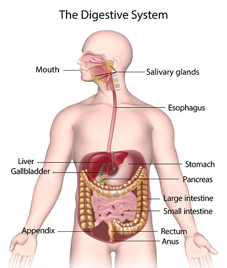
The function of the digestive system is to break down food into smaller and smaller compounds until it can be absorbed and used as energy.
It consists of a series of organs of the gastrointestinal tract and accessory digestive organs.
The organs of the digestive system encompass everything from the mouth to the anal canal, so it's actually one large conduit that includes the mouth, pharynx, esophagus, stomach, small intestine, and then the large intestine, ending at the anal canal.
The accessory digestive organs assist with the mechanical and chemical breakdown of food; these are the tongue, salivary glands, pancreas, liver, and gallbladder.
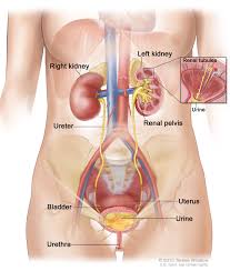
The urinary system is the body's drainage system. It consists of a group of organs that produce and excrete urine. These are the kidneys with their respective ureters, the urinary bladder, and the urethra.
The kidneys are a pair of retroperitoneal organs that remind us of a pair of beans. They have a rich blood supply that comes through the renal artery. The nephrons, inside the kidneys, filter the blood that passes through their network of capillaries (the glomerulus). This blood filtrate, or primary urine, then passes through a series of collecting tubules and ducts to eventually form the final ultrafiltrate, urine. The urine then enters the ureters, smooth muscle tubes that connect the kidneys to the urinary bladder. The bladder is a hollow, muscular organ that collects and stores urine before it is eliminated through urination. Some functions of the urinary system include: elimination of body waste products, regulation of blood volume and blood pressure, and regulation of electrolyte levels and blood pH.
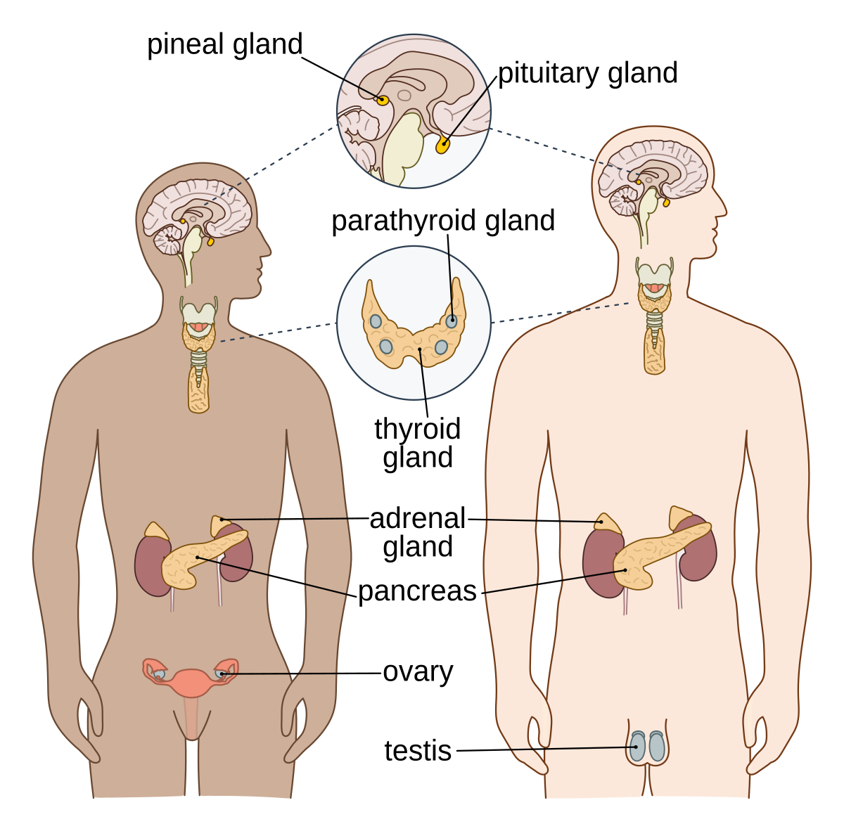
The endocrine system is a set of specialized organs (endocrine glands) distributed throughout the body that function by producing hormones.
Regarding the function of the endocrine system, the hormones it produces regulate a wide variety of bodily functions, such as triiodothyronine (T3), which regulates metabolism, and estrogen and progesterone, which regulate the menstrual cycle. Endocrine glands secrete hormones directly into the circulatory system to regulate the functions of remote target organs.
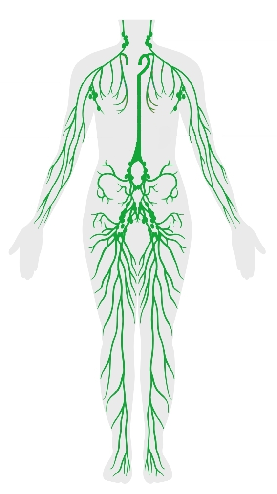
The lymphatic system is a network of lymphatic vessels that drain excess tissue fluid (lymph) accumulated in the intercellular compartments, filter it through the lymph nodes, expose it to lymphocytes (white blood cells) of the immune system, and then return the fluid to the circulatory system. The lymphatic system consists of lymph, lymphatic plexuses, lymphatic vessels, lymph nodes, and lymphoid organs. The function of this system is to transport and remove toxins and waste from our bodies; recirculate proteins; and defend us against invading microorganisms.
Lymph is a watery tissue fluid with a consistency similar to that of plasma. This begins as interstitial fluid that occupies the spaces between cells (intercellular space).
Excess fluid is collected by the lymphatic capillaries and transported through the lymphatic plexuses until it enters the lymphatic vessels, filtering through the lymph nodes along the way.
Superficial lymphatic vessels are found in the subcutaneous tissue next to the veins. These empty into the deep lymphatic vessels that travel alongside the arteries. The lymphatic vessels drain into the large lymphatic trunks, which join to form one of the two main collecting ducts: the thoracic duct and the right lymphatic duct.
The thoracic duct begins in the cisterna chyli. It collects lymph from the left side of the head, neck, thorax, left upper limb, abdomen, and both lower limbs, and then drains into the left venous angle (the junction of the left internal jugular vein and the left subclavian vein).
The right lymphatic duct drains the rest of the body and empties into the right venous angle. From the venous angles, the cleansed lymph is returned to the circulatory system and incorporated into the plasma. Previously, it was thought that the central nervous system did not have lymphatic vessels. However, more recent studies have found that its lymph is actually drained by lymphatic vessel-like structures found in the meninges.
The organs of the lymphatic system are divided into primary and secondary. The primary organs produce lymphocytes and release them into the lymphatic vessels. Two primary lymphoid organs are the thymus and the red bone marrow.
Some secondary lymphoid organs are the lymph nodes, tonsils, appendix, and spleen. Lymph nodes are masses of lymphoid tissue containing lymphocytes, attached to lymphatic vessels. The function of lymph nodes is to filter cellular debris, exogenous pathogens, excess tissue fluid, and leaking plasma proteins. There are some groups or chains of lymph nodes at key locations in our bodies (cervical, axillary, tracheal, inguinal, femoral, and para-aortic).
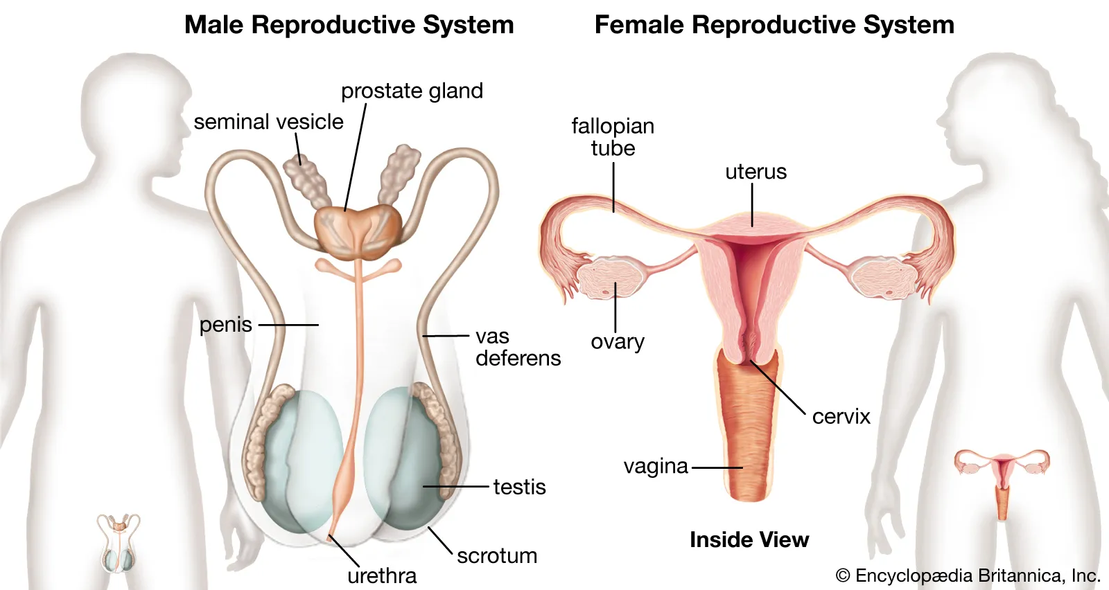
The reproductive system, or genital system, is a system of internal and external sexual organs that work together to contribute to the reproduction process. Unlike the other systems, the genital system has significant differences between the sexes.
The external female sexual organs, also known as the genitalia, are organs such as the vulva (the labia, clitoris, and vaginal opening). The internal sexual organs are the ovaries, uterine (fallopian) tubes, uterus, and vagina. The vulva provides protection and the entry point to the vagina and uterus, as well as adequate warmth and moisture to assist in sexual and reproductive functions. It is also important for arousal and orgasm in women.
The vagina is the canal that connects the outside of the body to the cervix. The ovaries secrete hormones and produce eggs, which are in turn transported to the uterus through the fallopian tubes. The uterus provides protection, nutrition, and waste disposal for the growing embryo or fetus. Additionally, contractions in the muscular wall of the uterus contribute to the expulsion of the fetus during childbirth.
The external male sex organs are the testes and penis, while the internal organs are the epididymides, vas deferens, and accessory glands. Functionally, they can be grouped into three categories.
The first is for sperm production (the testes) and sperm storage (the epididymides). The second category of organs produces ejaculatory fluid; the vas deferens and the accessory glands (the seminal vesicles and prostate). The last category is responsible for copulation and sperm deposition; these organs are the penis, urethra, and vas deferens.
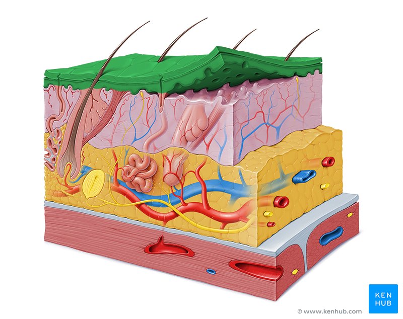
The integumentary system is a group of organs that form the outer covering of the body. This includes the skin and its appendages, sweat glands, and sensory receptors.
The skin is the largest organ in the human body. It has three layers: the epidermis, dermis, and hypodermis.
The epidermis is a thick, keratinized epithelium composed of multiple layers.
Beneath the epidermis is the dermis, a layer of connective tissue that contains the blood vessels and nerves that provide vascularization and innervation to the skin.
The underlying fascia, also called the hypodermis, consists of fat, connective tissue, and skin appendages (hair, nails, sebaceous and sweat glands). The functions of the integumentary system are diverse. It forms a continuous layer that protects the body from harmful events, such as external injuries, water and heat loss, and the carcinogenic effects of ultraviolet radiation. It also excretes some waste, contains pain, sensation, pressure, and temperature receptors, and synthesizes vitamin D.
Remember to check the answers to the open-ended questions at the bottom of this page.
Once you click this button, the questions will close and you will not be able to change your answer.
We recommend visiting the following material for further knowledge or understanding on the topic:
1. Human body systems 2. Human Body 3. Functions of All 11 Organ Systems in the Human Body6. Groups of organs that work together to produce and maintain vital functions.
7. The spinal cord transmits signals between the brain and the rest of the body and is also responsible for some quick, involuntary reflexes.
8. The digestive system includes the gastrointestinal tract (mouth, pharynx, esophagus, stomach, small intestine, large intestine, anal canal) and accessory organs such as the pancreas, liver, gallbladder, and salivary glands.
9. The integumentary system protects the body from external damage, regulates temperature, excretes waste, contains sensory receptors, and synthesizes vitamin D.
10. The skeletal system is divided into the axial skeleton, which includes the bones of the head and trunk, and the appendicular skeleton, which includes the bones of the limbs and the shoulder and pelvic girdles.
References:
1. Sistemas del cuerpo humano. (2023, 30 octubre). Kenhub. https://www.kenhub.com/es/library/anatomia-es/sistemas-del-cuerpo-humano
2. Human body systems. (2023, november 3). Kenhub. https://www.kenhub.com/en/library/anatomy/human-body-systems
3. The Editors of Encyclopaedia Britannica. (2025, april 21). Human body | Organs, Systems, Structure, Diagram, & Facts. Encyclopedia Britannica. https://www.britannica.com/science/human-body
4. Narayana Health. (w.d.). Narayana Health. https://www.narayanahealth.org/blog/organ-systems-all-11-and-what-they-do
5. Amoeba Sisters. (2024, 23 febrero). Human body Systems Overview (Updated 2024) [Vídeo]. YouTube. https://www.youtube.com/watch?v=0JDCViWGn-0
6. National Geographic. (2017, 1 diciembre). Human Body 101 | National Geographic [Vídeo]. YouTube. https://www.youtube.com/watch?v=Ae4MadKPJC0
7. MooMooMath and Science. (2022, 13 julio). 11 Body Systems in 3 minutes [Vídeo]. YouTube. https://www.youtube.com/watch?v=Cpb0RBBL9Wc